Failure Analysis
To solve a problem, you first have to identify it.
We are an independent analysis laboratory for battery technology, focused on detailed failure analysis of complete systems and battery cells.
Our analysis team finds every defect, from a faulty weld on the housing to a particle inside the cell, using the latest technologies such as computed tomography and scanning electron microscopy.
On the trail of every fault
We examine every component of a complete battery, whatever its condition, including after field incidents or destructive tests. Even where the damage is severe, the right process can still trace the causes and support important decisions. With our broad range of analysis methods we can locate and investigate any defect. Our systematic teardown documents the chain of evidence at every stage of disassembly.
Reading the BMS fault memory via CAN
Electrical evaluation
Non-destructive analysis
- 3D laser scanning for complete dimensional measurement
- Pressure testing of housings and cooling circuits
- High-precision helium leak testing
Battery disassembly
- Recovery of damaged batteries after abuse tests and field incidents
- Safe discharge of damaged HV batteries
- Controlled disassembly of the complete battery down to component level
- Failure analysis of the complete battery including all sub-components
- Detailed photo documentation throughout the disassembly process
Combined applications
High-precision voltage measurements in a climate-controlled environment let us identify the problem cell, from module through cell pack down to single-cell level. A preparation method developed in-house then narrows the problem down to the individual single cell. A CT scan of the suspect cell can reveal the type of defect, and the teardown analysis finds the defect and documents it in detail. The laser microscope, the scanning electron microscope or the grinding and polishing machine can then analyse the anomalies further, so that the origin of the problem can be traced.
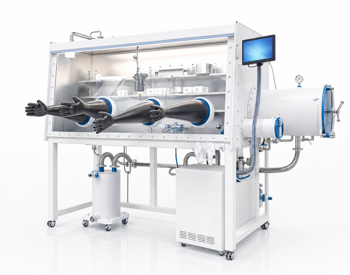
An RPT identifies the faulty cells, and a CT scan of those cells can reveal the cause of the capacity problem. A teardown analysis at 100% SOC shows the intercalation behaviour of each anode. Safely opening a fully charged lithium-ion cell requires an inert atmosphere: our argon glovebox keeps O2 and H2O below 10 ppm and preserves the electrode chemistry, so that defects can be attributed unambiguously.
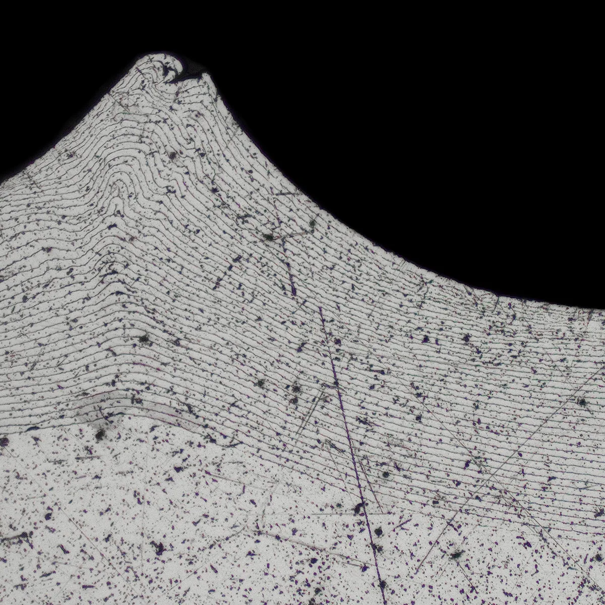
Weld quality is a decisive reliability factor in battery systems, at battery, module and cell level alike. A CT scan captures cracks as well as pore size and distribution. A metallographic section shows the weld penetration depth and the bonded area.
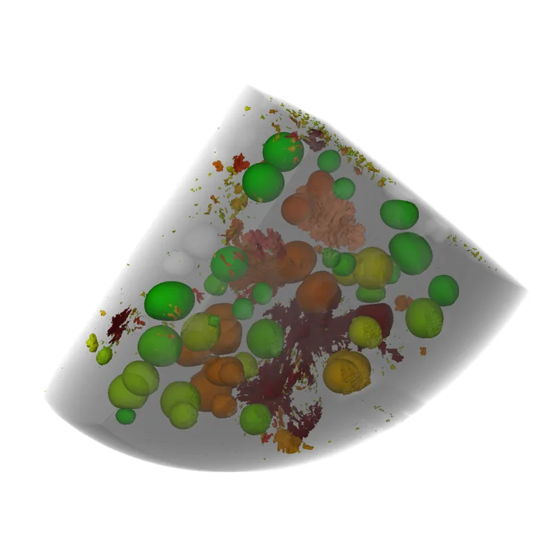
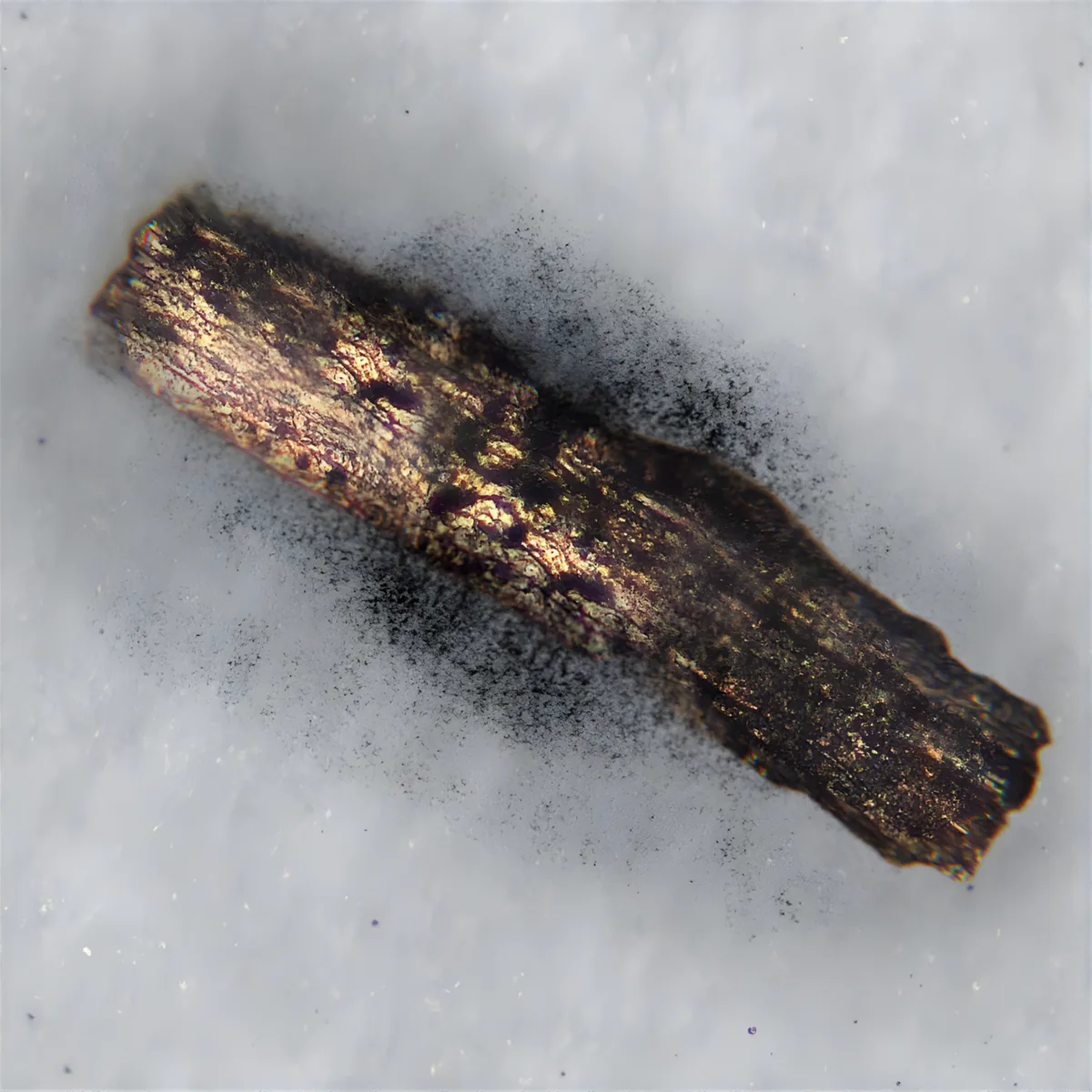
Because battery cell manufacturing is so complex, particles can enter the cell at many different points in production. Even the smallest particle, in the wrong place inside the cell, can have drastic consequences for the cell and the complete battery. That makes it all the more important to analyse the particles found in detail, to establish at which process step the contamination occurred.
In our cycling chambers, the ageing of a cell or module can be reproduced safely with defined charge and discharge cycles at a defined temperature. A subsequent teardown analysis documents every sign of ageing in detail.
Which fault will we find next?
With our advanced laboratory equipment, no failure pattern stays hidden.



Can't find the service you need? Contact us. Our laboratory is versatile and flexible and can respond to individual requirements.
Can battery modules and cells be CT scanned before teardown, even at a high state of charge?
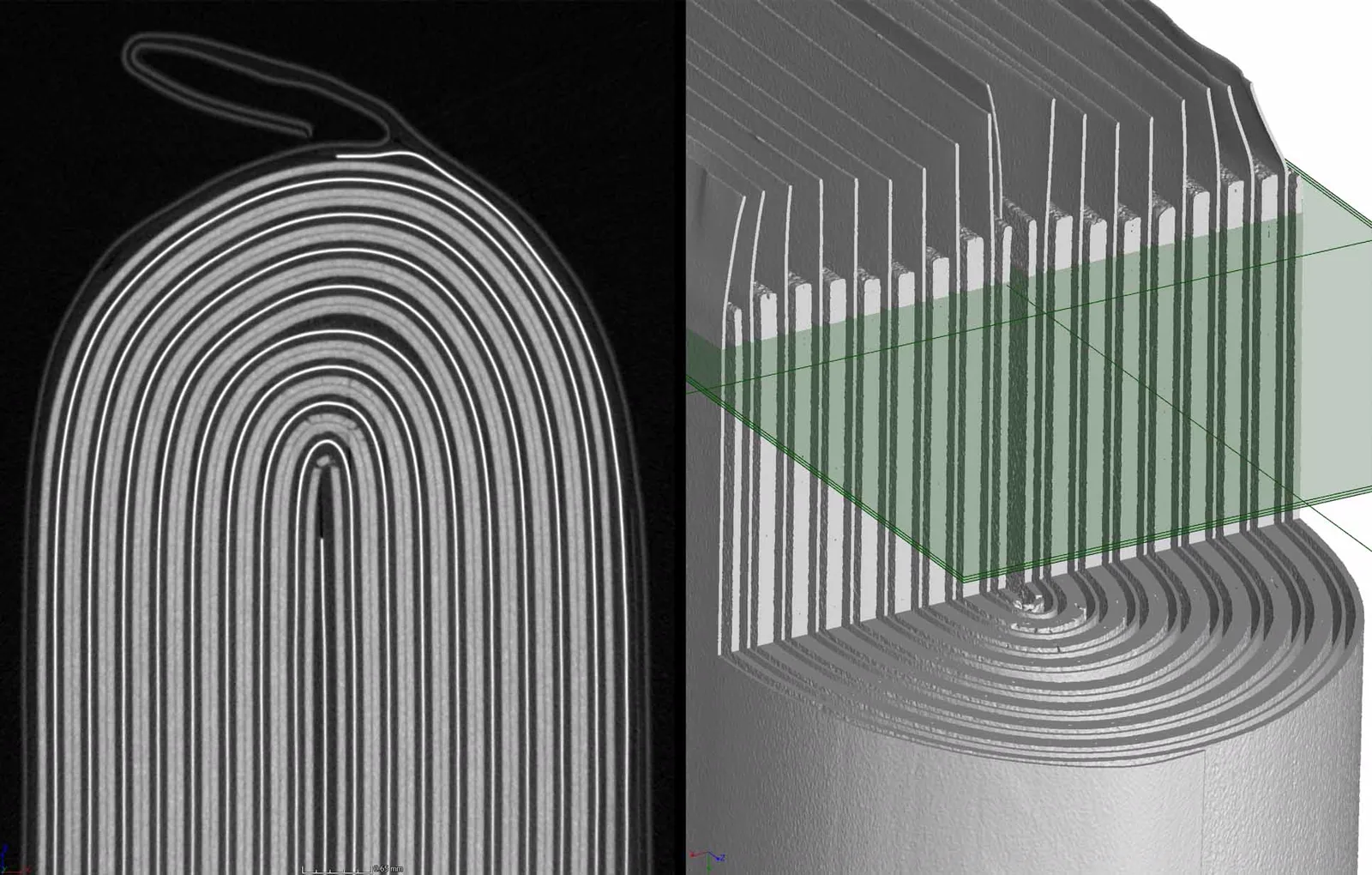
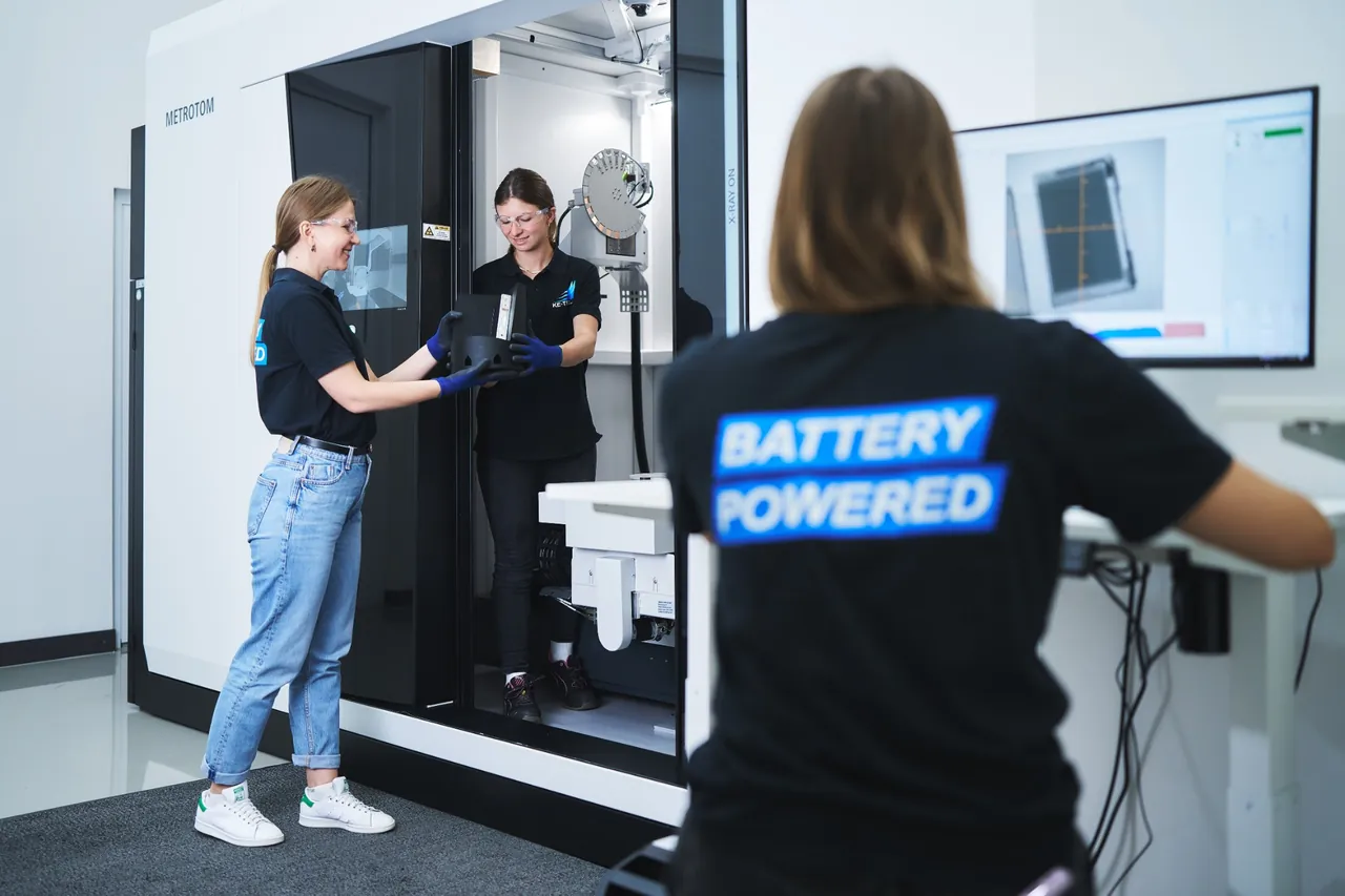
Yes, our CT scanner is also used on battery modules and cells at 100% SOC. This confirms a fault inside the specimen before the disassembly analysis begins. The Zeiss Metrotom 1500 works to VDI/VDE 2630-1.3 and takes specimens up to 870 mm high, 615 mm in diameter and 50 kg, so complete EV modules as well as single cells.
What defects can be found in a cell?
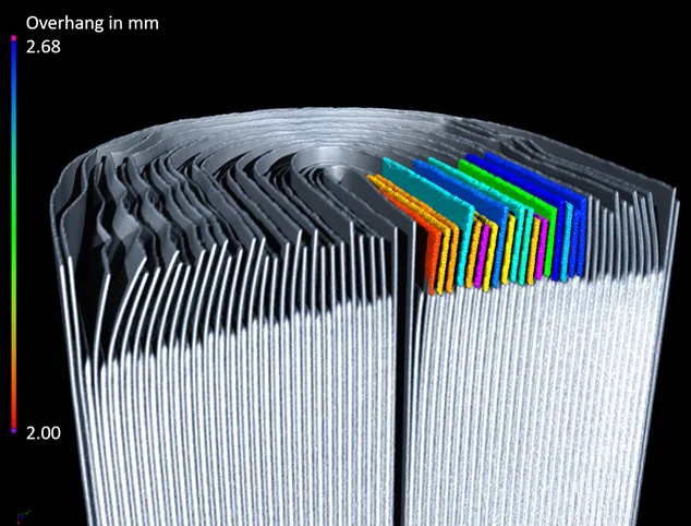
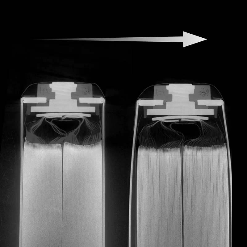
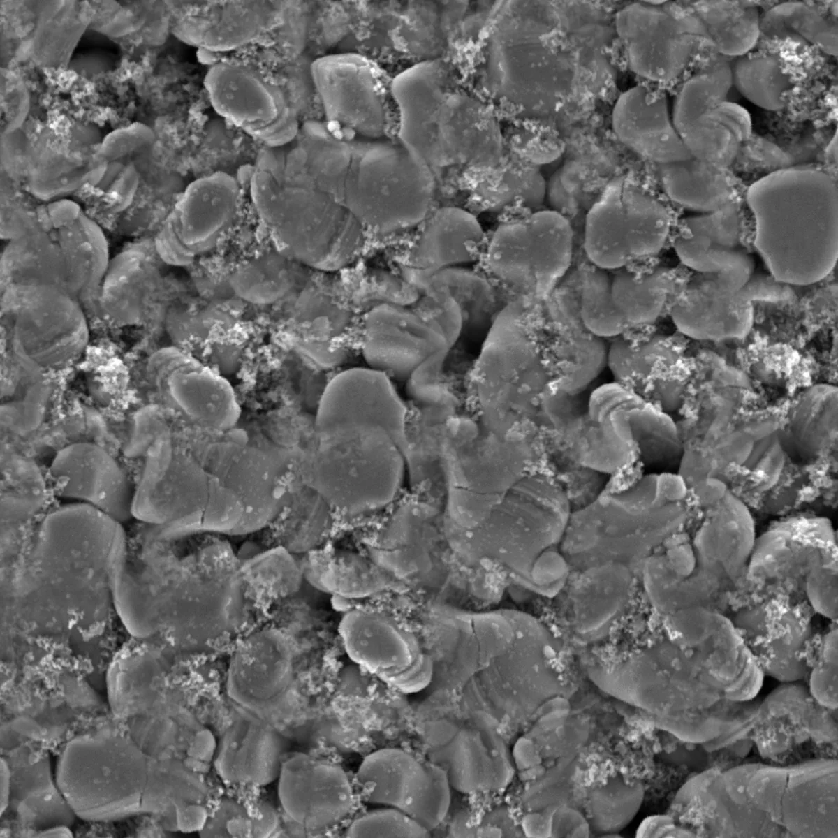
We detect, for example, metallic and non-metallic foreign particles, voids and pores in welded joints, folded electrodes, and sharp cut edges and micro-holes in the separator. Add to that collapsed windings in cylindrical cells (core collapse), which can pierce the separator, and deposits from unwanted side reactions inside the cell. Elemental analysis also tells us what any foreign bodies are made of, so process faults can be traced back conclusively. Our experienced analysis team knows the problems battery cells have, recognises them, and applies analysis methods in a targeted way to gather all the information needed about the defect.
How do you stop a teardown from distorting the analysis?
Our cell laboratory and analysis team specialise in examining specimens as cleanly as possible, so that nothing influences the result. The specimen is photographed at every disassembly step, so no information is lost.
Can you analyse single cells only, or complete batteries too?
We have the methodological expertise for a safe disassembly process across a complete EV battery down to the single cell, damaged or not.
Why should a cell be examined non-destructively before it is opened?
Disassembly analyses are very laborious and cannot be undone. CT scans let us see inside a cell without opening it and find manufacturing problems, foreign particles or material defects. That makes it possible to choose in advance which cells are worth disassembling, and the scan also provides excellent traceability.
How do you decide which analyses are needed to find the root cause?
Our experienced team will gladly advise you on which combination of methods makes sense for your particular case.
Can critical cells be analysed too?
Yes, our wide range of disassembly methods lets us take cells apart in any condition.
Can solid-state cells be analysed too?
Yes, our analysis team also has experience disassembling solid-state cells with pure lithium anodes.
Your question isn't covered? Contact us. Our laboratory is versatile and flexible and can respond to individual requirements.
Whether troubleshooting a complete battery or going straight to a single cell, our analysis team helps you identify any problem.